Foot Muscles Mri : Mri Of The Ankle Detailed Anatomy W Radiology. Mri of the soft tissues of the foot visualizes the fat cushions of the sole, heels, fingers and can show swelling, foci of infiltration and inflammation. Those fibers of the most medial and largest belly are known as. The aim of this study is to describe clinical and mri patterns of … Mri and ultrasound have been utilised in the assessment of the plantar intrinsic foot muscles. Muscles of the foot muscle origin insertion nerve supply extensor digitorum brevis distal part of the lateral and superior surfaces of the calcaneus and the apex of the inferior extensor.
The majority of soft tissue lesions in the foot and ankle are benign. The aim of this review is to provide the reader with a comprehensive overview of the magnetic resonance imaging (mri) characteristics of the most common benign and malignant soft tissue neoplasms which occur around the foot and ankle. 6 mri is commonly ordered in the diabetic patient to rule out infection in the presence of an ulcer, to evaluate the severity of charcot arthropathy. Distal part of the lateral and superior surfaces of the calcaneus and the apex of the inferior extensor retinaculum. Mri is an ideal method for identifying areas of muscle atrophy and fatty infiltration.
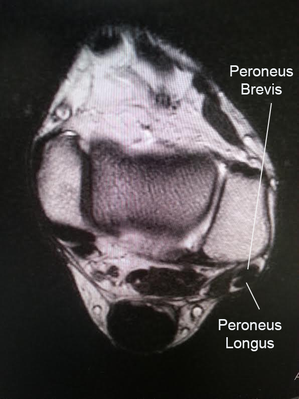
Ankle Tendons Topographic Anatomy Radiology Case Radiopaedia Org from prod-images-static.radiopaedia.org Electromyography in cases of foot drop involves testing of the tibialis anterior as well as muscles innervated by the superficial peroneal, tibial, sciatic, and superior gluteal nerves. Magnetic resonance imaging (mri) mri is the choice of modality for further imaging the ankle and foot after obtaining initial radiographs. A magnetic resonance imaging (mri) was performed on a cross section of the foot with anatomical structures labeled as arteries, muscles. General anatomy and the musculoskeletal system: Mri and ultrasound have been utilised in the assessment of the plantar intrinsic foot muscles. Magnetic resonance imaging, otherwise known as mri, uses a combination of magnetic fields and radio waves to take images of the internal structures of your body. The paraspinal muscles, which are innervated by the spinal nerve dorsal ramus, are also frequently tested. The purpose of this study was to examine the muscle functional (mf) mri and emg responses to perturbations of the foot by running in varus, neutral and valgus wedged shoes.
6 mri is commonly ordered in the diabetic patient to rule out infection in the presence of an ulcer, to evaluate the severity of charcot arthropathy.
The adductor hallucis has two heads: Mri and ultrasound have been utilised in the assessment of the plantar intrinsic foot muscles. Both muscles are innervated by the deep fibular nerve. General anatomy and the musculoskeletal system: A magnetic resonance imaging (mri) was performed on a cross section of the foot with anatomical structures labeled as arteries, muscles. Mri has surpassed nuclear medicine imaging due to the greater specificity of mri and its ability to delineate osseous anatomy as well as discrete abscesses and sinus tracts diagnostic of infection. The flexor digitorum brevis muscle lies immediately superior to the plantar aponeurosis and inferior to the tendons of the flexor digitorum longus in the sole of the foot. Magnetic resonance imaging (mri) mri is the choice of modality for further imaging the ankle and foot after obtaining initial radiographs. Electromyography in cases of foot drop involves testing of the tibialis anterior as well as muscles innervated by the superficial peroneal, tibial, sciatic, and superior gluteal nerves. Plantar interossei (foot) dr yuranga weerakkody ◉ and dr geon oh et al. In addition, an image of all the muscles of the back and plantar part of the foot, all tendons and tendon ligaments, blood vessels and nerves are obtained. Coronal images are perpendicular to the long axis of the metatarsals. Related posts of foot muscle anatomy mri muscle anatomy knee mri.
The interosseous muscles of the foot are muscles found near the metatarsal bones that help to control the toes. The flexor digitorum brevis muscle lies immediately superior to the plantar aponeurosis and inferior to the tendons of the flexor digitorum longus in the sole of the foot. 6 mri is commonly ordered in the diabetic patient to rule out infection in the presence of an ulcer, to evaluate the severity of charcot arthropathy. The purpose of this study was to examine the muscle functional (mf) mri and emg responses to perturbations of the foot by running in varus, neutral and valgus wedged shoes. Routine ankle magnetic resonance imaging (mri) tests involve taking images of the foot and ankle in the axial, coronal, and sagittal planes parallel to the tabletop(2).
Peroneal Tendon Tears And Instability Foot Ankle Orthobullets from upload.orthobullets.com Swelling and tenderness in your joints. Mri is an ideal method for identifying areas of muscle atrophy and fatty infiltration. Mri with user outlined plantar intrinsic and extrinsic muscles group. The aim of this study is to describe clinical and mri patterns of … Routine ankle magnetic resonance imaging (mri) tests involve taking images of the foot and ankle in the axial, coronal, and sagittal planes parallel to the tabletop(2). As the fiber bundles extend distally, they become grouped into four bellies. Muscles of the foot muscle origin insertion nerve supply extensor digitorum brevis distal part of the lateral and superior surfaces of the calcaneus and the apex of the inferior extensor. Magnetic resonance imaging (mri) is the modality of choice in diagnosing accessory muscles, delineating their relationship to adjacent structures, and differentiating them from soft tissue tumors.
Also known as osteomyelitis, which is generally treated with antibiotics, but can lead to an amputation.
The interosseous muscles of the foot are muscles found near the metatarsal bones that help to control the toes. Adductor hallucis is anatomically located in the central compartment of foot, but the muscle is functionally grouped with the medial plantar muscles of foot because it acts on the great toe (hallux). Plantar interossei (foot) dr yuranga weerakkody ◉ and dr geon oh et al. Coronal images are perpendicular to the long axis of the metatarsals. They are considered voluntary muscles. 6 mri is commonly ordered in the diabetic patient to rule out infection in the presence of an ulcer, to evaluate the severity of charcot arthropathy. The adductor hallucis has two heads: Magnetic resonance imaging, otherwise known as mri, uses a combination of magnetic fields and radio waves to take images of the internal structures of your body. The three plantar interossei muscles adduct the 3 rd, 4 th and 5 th toes toward the long axis through the 2 nd toe. This imaging technique assesses the ligaments and tendons, neurovascular structures ( tarsal tunnel and plantar fascia), and the osseous structures (19). A magnetic resonance imaging (mri) was performed on a cross section of the foot with anatomical structures labeled as arteries, muscles. Foot and (from schuenke m, schulte e. The purpose of this study was to examine the muscle functional (mf) mri and emg responses to perturbations of the foot by running in varus, neutral and valgus wedged shoes.
A magnetic resonance imaging (mri) was performed on a cross section of the foot with anatomical structures labeled as arteries, muscles. They are considered voluntary muscles. The interosseous muscles of the foot are muscles found near the metatarsal bones that help to control the toes. Your doctor, with the help of a radiologist, can then examine these images to determine whether there is anything wrong with your foot or ankle. Related posts of foot muscle anatomy mri muscle anatomy knee mri.
Foot Mri Anatomy Anatomy Drawing Diagram from i.pinimg.com Lateral and medial processes of calcaneal tuberosity. Ten males ran at 3.6 m/s in specially constructed shoes for 5 min with. Mri has surpassed nuclear medicine imaging due to the greater specificity of mri and its ability to delineate osseous anatomy as well as discrete abscesses and sinus tracts diagnostic of infection. Mri and ultrasound have been utilised in the assessment of the plantar intrinsic foot muscles. As the fiber bundles extend distally, they become grouped into four bellies. The aim of this study is to describe clinical and mri patterns of … The purpose of this study was to examine the muscle functional (mf) mri and emg responses to perturbations of the foot by running in varus, neutral and valgus wedged shoes. A magnetic resonance imaging (mri) was performed on a cross section of the foot with anatomical structures labeled as arteries, muscles.
Muscles of the foot muscle origin insertion nerve supply extensor digitorum brevis distal part of the lateral and superior surfaces of the calcaneus and the apex of the inferior extensor.
The interosseous muscles of the foot are muscles found near the metatarsal bones that help to control the toes. The paraspinal muscles, which are innervated by the spinal nerve dorsal ramus, are also frequently tested. Mri has surpassed nuclear medicine imaging due to the greater specificity of mri and its ability to delineate osseous anatomy as well as discrete abscesses and sinus tracts diagnostic of infection. Mri with user outlined plantar intrinsic and extrinsic muscles group. • muscle edema is seen secondary to multiple etiologies including trauma, infectious and inflammatory processes, autoimmune disorders, neoplasms, and denervation injuries • on mri muscle edema is characterized by increase in free water within the muscle • muscle edema is seen on mri as increased signal on fluid sensitive sequences t2 fs Related posts of foot muscle anatomy mri muscle anatomy knee mri. Electromyography in cases of foot drop involves testing of the tibialis anterior as well as muscles innervated by the superficial peroneal, tibial, sciatic, and superior gluteal nerves. This imaging technique assesses the ligaments and tendons, neurovascular structures ( tarsal tunnel and plantar fascia), and the osseous structures (19). Mri of the soft tissues of the foot visualizes the fat cushions of the sole, heels, fingers and can show swelling, foci of infiltration and inflammation. Both muscles are innervated by the deep fibular nerve. Magnetic resonance imaging, otherwise known as mri, uses a combination of magnetic fields and radio waves to take images of the internal structures of your body. Coronal images are perpendicular to the long axis of the metatarsals. The three plantar interossei muscles adduct the 3 rd, 4 th and 5 th toes toward the long axis through the 2 nd toe.
Share this post
0 Response to "Foot Muscles Mri : Mri Of The Ankle Detailed Anatomy W Radiology"


0 Response to "Foot Muscles Mri : Mri Of The Ankle Detailed Anatomy W Radiology"
Posting Komentar